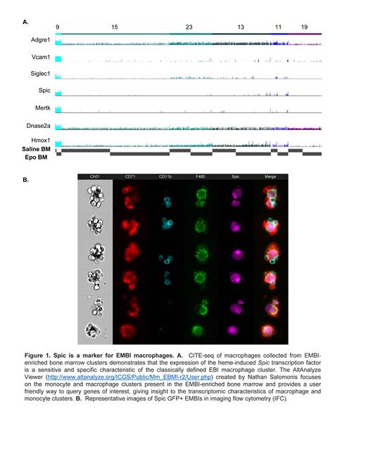
We recently demonstrated that the majority of the erythroblastic islands in the mouse bone marrow are supporting not only terminal erythropoiesis but also granulopoiesis, being common niches for terminal hematopoiesis (Romano, Seu, et al. Blood 2022). Using single cell RNA-sequencing (scRNA-seq) and Cellular Indexing of Transcriptome and Epitopes by Sequencing (CITE-seq) we demonstrated several macrophage populations captured from EMBI-enriched bone marrow. Notably, the population of the classically defined EBI macrophages characterized by the genes Mertk, Dnase2a, and Hmox1, also showed sensitive and specific expression of the heme-induced Spic transcription factor (Figure 1). In agreement with the CITE-seq data, imaging flow cytometry (IFC) of EMBIs from SpiC igfp/igfp mouse bone marrow demonstrated that at least 80% of the central macrophages were GFP +, confirming their expression of Spic (Figure 2). After stimulation with erythropoietin (Epo), which leads to an increase of both the number and size of EMBIs, both Spic-GFP-positive and negative central macrophages doubled in number, as determined by IFC studies. Moreover, Spic-GFP + EMBI macrophages isolated from mouse bone marrow reconstituted efficiently into EMBIs after incubation with CD71 + and CD11b + cells isolated from another ( Spic-GFP neg) bone marrow.
Characterizing EMBIs by IFC allows for identification of phenotypic characteristics of macrophages participating in the EMBI structure. The strong and intimate interactions between the erythroblasts and granulocytic precursors to the central macrophage facilitates the preservation of EMBIs and their analysis by IFC; however, these same strong interactions add a layer of complexity to methods that require single cells, such as scRNA-seq. Previous attempts to capture EMBI macrophages have had lower than expected yields. Utilizing the Spic-GFP reporter mouse model permitted the use of harsher dissociation by trypsinization and sorting based on GFP positivity, optimizing EMBI macrophage recovery. The resulting captures yielded thousands of EMBI macrophages based on cells expressing Adgre1, Vcam1, Hmox1, DNase2a, and Mertk that show a marked degree of transcriptional heterogeneity in other macrophage surface markers. Comparison of the captures from control (saline-) and Epo-stimulated EMBI macrophages revealed an emergent macrophage population, increasing up to 25-fold after Epo in comparison to control. This Epo-induced macrophage population has approximately 900 globally distinguishing genes including markers characteristic of monocytes such as Ace (encoding the angiotensin-converting enzyme), implicating monocyte recruitment to serve as stress erythropoiesis-supporting macrophages within the murine bone marrow. Additional clusters of macrophages with the characteristics of classically defined EMBI macrophages are also increasing with Epo- stimulation, indicating heterogeneity within the Spic-GFP + population. To verify these transcriptomic results on the protein level, we performed IFC with CD169, Mertk, and other EMBI macrophage markers providing further insights to the heterogeneity of their expression on the central macrophages within intact EMBIs. We anticipate that the Spic-GFP + reporter mouse will be a useful tool in EMBI studies, providing a specific and sensitive marker to trace and isolate the majority of EMBI macrophages.
Disclosures
Kalfa:Agios Pharmaceuticals, Inc.: Consultancy, Research Funding; Forma/Novo Nordisk: Consultancy, Research Funding.


This feature is available to Subscribers Only
Sign In or Create an Account Close Modal